I've been diagnosed
Start with what happens next
A diagnosis is a lot to take in. You do not need to understand everything today. Here is the shape of the journey, so you can start where you want to start.
Reason for hope
Found early and treated, breast cancer has a high chance of recovery. A team of specialists will guide each step with you.
Your care team
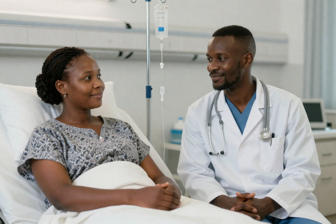
Breast cancer is treated by a multidisciplinary team, so several specialists plan your care together.
Each person on that team is an expert in their own field. You are the expert on you. Your questions, your worries and what matters to you belong in the plan, so say them out loud.
A surgeon and a cancer doctor (oncologist)
A radiologist and a radiographer for scans and radiation
A pathologist who examines the tissue
A specialist nurse, and a reconstructive surgeon if needed
Physiotherapy, dietetics, and psychology support
You can ask for a second opinion
Asking another doctor to look at your diagnosis is normal, and you are allowed to do it. It can confirm what you have been told, or it can open up other treatment options to talk through with your own doctor.
Questions to ask your doctor
Take these with you, and write the answers down.
- What type of breast cancer do I have, and what stage is it?
- What are my treatment options?
- Can I have a second opinion?
- What side effects should I expect, and how are they managed?
- Where can I get support for how I am feeling?
Types of breast cancer
There are several types. Your team will tell you which one you have, which guides treatment.
Ductal carcinoma in situ. Abnormal cells are still contained inside a milk duct and have not spread. It is non-invasive.
Types are also grouped by what drives them: hormone-receptor positive, HER2 positive, or triple negative.
Special situations
Breast cancer can affect some groups differently. Fuller guides for each are coming.
Breast cancer in men
Men have a small amount of breast tissue and can get breast cancer too, though it is rare. Any lump or change behind the nipple should be checked.
Younger women
Breast cancer is less common before 40, but it does happen and can grow faster. Knowing your normal and acting on changes matters at any age.
During pregnancy
Breast cancer is rare during pregnancy and can still be treated. A care team plans treatment to protect both mother and baby. A fuller guide is coming.
How it is diagnosed
Diagnosis usually moves through these steps.
Clinical breast examination
A doctor or nurse feels both breasts and the armpits for any lump or change.
Mammogram and ultrasound
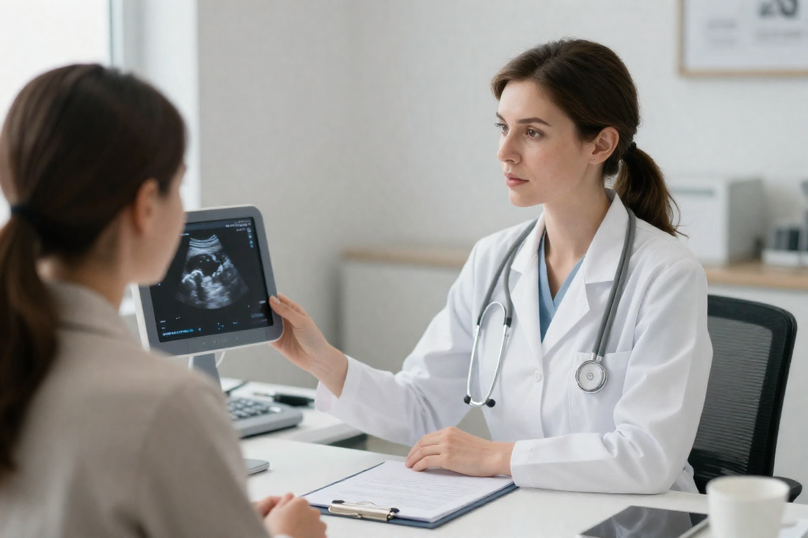
A mammogram is a low-dose breast x-ray. An ultrasound uses sound waves and can show whether a lump is solid or a fluid-filled cyst.
Biopsy
A small sample of tissue or cells is taken and examined under a microscope. For most breast cancers, a biopsy is the only way to be sure.
Further tests
An MRI, blood tests, or genetic and biomarker tests may be done to plan the right treatment.
Understanding your results
Your results describe the cancer so the team can match treatment to it.
Grade
How much the cells look like normal cells. Grade 1 cells look near-normal and grow slowly. Grade 3 cells look abnormal and can grow faster.
Tumour
Its size, where it is, and how many tumours there are.
Lymph nodes
Whether cancer has reached the lymph nodes (node-positive) or not (node-negative).
Receptors (ER, PR, HER2)
Your cells are tested for two hormone receptors, oestrogen (ER) and progesterone (PR), and for a protein called HER2. These three decide the type of breast cancer you have and which treatments will work, so this is one of the most important parts of your result.
Stages, from 0 to 4
The stage describes how far the cancer has spread.
Stage 0
Still in its place of origin and not spread to nearby tissue (in situ).
You may also hear the TNM system, which scores the tumour (T), the lymph nodes (N), and any spread or metastasis (M).
Treatment options
Most people have a combination, planned by the team for their type and stage.
Local treatment (the breast itself)
Surgery, breast-conserving
Removing the tumour and a little surrounding tissue, such as a lumpectomy or wide local excision.
Surgery, mastectomy
Removing all the breast tissue, sometimes with lymph nodes. Reconstruction may be an option.
Radiotherapy
Targeted radiation to kill cancer cells or stop them growing, while sparing healthy tissue as much as possible.
Systemic treatment (the whole body)
Chemotherapy
Medicines that kill cancer cells or stop them dividing.
Hormone therapy
Used for cancers that are sensitive to oestrogen or progesterone.
Targeted therapy
Newer medicines that work differently from chemotherapy, aimed at specific features of the cancer.
Immunotherapy may be used in some cases. Detailed, reviewed guidance is being added.
Managing side effects
Knowing what to expect, and that side effects can be managed, is one of the most valued parts of this journey.
Treatment brings side effects, and most of them can be managed. Tell your team about anything you notice, even if it feels small or silly.
Common ones are: tiredness that rest does not fix, pain, hair and skin changes, going into menopause early, trouble with memory and concentration (often called chemo brain), and effects on your bones and heart that your team will watch.
None of this is something you have to put up with quietly. There is almost always something that helps.
Newly diagnosed? You are not alone.
CANSA's toll-free help line can talk you through what is next, in your own language.